
Have you ever wondered how clinicians look past routine jaw discomfort to uncover complex temporomandibular joint (TMJ) disorders? While CBCT (Cone Beam Computed Tomography) is traditionally celebrated for mapping bony structures, a recent case in our practice highlights how dynamic positioning—specifically the open-mouth view—can offer incredible diagnostic clues about soft tissue issues like disc displacement.
Let’s dive into an intriguing clinical case and explore why the open-mouth position is often far more revealing than the closed-mouth view.
The Case: An 18-Year-Old Male Presenting with Severe Restricted Opening
Recently, we got a CBCT for reporting, where we observed a patient complaining of severe pain and noticeable restriction whenever he tried to open his mouth. Because clinical evaluation pointed toward a significant internal issue, a CBCT scan was advised to examine the underlying bony architecture of the temporomandibular joints.
Upon clinical examination, we noted a severely restricted mouth opening accompanied by a noticeable deviation toward the right side. Initially, we suspected impacted third molars—a frequent culprit in patients of this age group—prompting a CBCT scan to rule out dental causes and properly evaluate the temporomandibular joints.
When we reviewed the scans, the presentation was fascinating. While closed-mouth views provide a static baseline, the open-mouth position proved vastly superior in capturing functional asymmetry and joint dynamics.
Key CBCT Findings
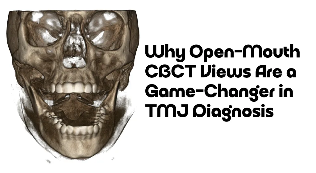
Left Condyle Pathologies: The scan revealed distinct osteolytic areas accompanied by subchondral osteosclerosis in the left condyle, alongside clear erosive changes signaling active degenerative joint disease.
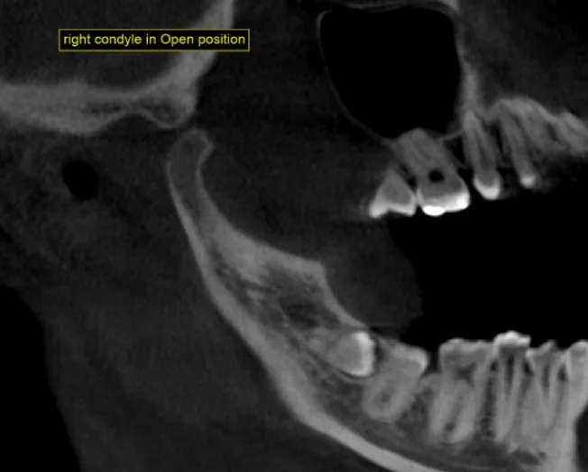
Asymmetrical Condylar Movement: The open-mouth position vividly demonstrated normal translational movement of the right condyle, contrasted against severely restricted movement on the left side.
Dental Impactions & Angulations: Incidental findings included a severely mesially inclined (almost horizontal) impacted tooth #48 whose crown was impinging on the distal surface of #47, as well as mesial and lingual inclination of tooth #38.
Why Open-Mouth CBCT Positions Outshine Closed-Mouth Views
In routine diagnostics, practitioners often rely heavily on closed-mouth CBCT imaging. However, dynamic imaging (capturing the joint in motion) changes everything:
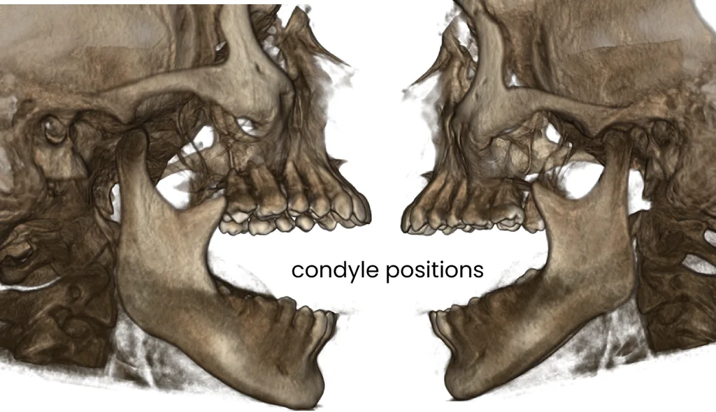
Unmasking Functional Restriction: A closed-mouth view only shows static positioning. The open-mouth view allows clinicians to directly observe translation and symmetry, instantly highlighting hypomobility or mechanical blocks on the affected side.
Clues to Soft Tissue Pathology: Although CBCT is fundamentally a bone-imaging modality, observing how the condyle fails to translate past the articular eminence provides strong secondary clues. In this patient, the restricted movement combined with degenerative erosion strongly mirrors clinical presentations of disc displacement without reduction (closed lock).
Visualizing Degenerative Changes Under Stress: Erosive changes in the condyle become clinically significant when observed under functional loading, helping differentiate simple muscle-related jaw fatigue from true structural joint pathology.
Decoding the Disc: How Bone Imaging Hints at Soft Tissue Trouble
While magnetic resonance imaging (MRI) remains the gold standard for directly visualizing the articular disc, our CBCT findings provide powerful indirect evidence of disc pathology.
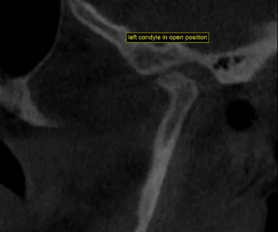
When closely examining the left joint, a few critical details demand our attention. First, notice the significantly narrowed joint space in the left condyle—a classic indicator of disc collapse causing the condyle to seat too closely against the glenoid fossa. Don’t be confused by the condyle’s apparent inferior positioning, where there seems to be a large gap between the glenoid fossa and the condyle; this is simply a natural byproduct of the open-mouth translation, whereas the actual articulating surface is located antero-superiorly, where we can see near-contact with the bone (Level 3 Anterior disc displacement).
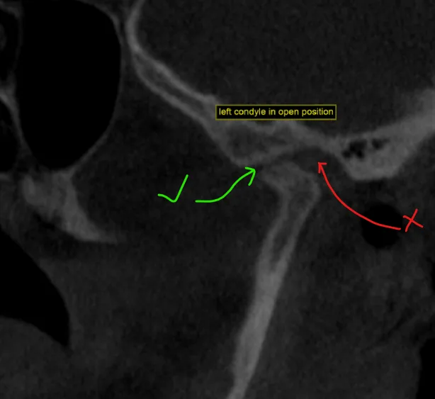
Second, observe the severe erosive changes on the condyle’s articular surface, which clearly point to elevated mechanical friction and ongoing degenerative stress within the joint.
Moreover, the severely restricted mouth opening on the left side, combined with the lack of condylar translation and the presence of subchondral erosions, strongly points toward disc displacement without reduction (commonly known as a closed lock). When the disc is permanently displaced anteriorly without reducing back into place, it acts as a mechanical physical barrier. This prevents the left condyle from sliding forward smoothly onto the articular eminence, forcing the mandible to deviate and causing the patient excruciating pain during translation.
The Takeaway
Isn’t it fascinating how a diagnostic tool built for bone can help us piece together the puzzle of soft tissue dysfunction? By utilizing dynamic open-mouth CBCT views, clinicians can move beyond static imaging and observe the true biomechanical story of the TMJ.
Have you or your patients ever experienced unexplained jaw locking or restricted opening? How has advanced imaging changed your perspective on dental and joint diagnostics?

A passionate dental educator with more than 13 years of academic experience, dedicated to making complex imaging feel simple by starting from the basics. Technology is his playground—specializing in CBCT reporting, digital implantology, 3D modelling, and dental 3D printing innovations. Through his writing and teaching, he loves empowering clinicians to bridge the gap between advanced digital workflows and everyday clinical practice.
Read more about me on the “about me” page.
- Decoding TMJ Pain: Why Open-Mouth CBCT Views Are a Game-Changer in TMJ Diagnosis - October 30, 2026
- What is Dental MRI? The Future of Radiation-Free Oral Imaging - October 25, 2026
- The Dual-Kit Dilemma: Why Fragmented Guided Implant Systems Are Losing Ground to Single-Kit Solutions - October 24, 2026