Gone are the days when dental diagnostics relied solely on 2D radiographs to identify pathologies. While traditional panoramic (OPG) and periapical radiographs offered invaluable glimpses into dental anatomy, they often fell short in accuracy and detail. Today, diagnostic 3D modelling—such as CT or CBCT scans—delivers unparalleled perfection, allowing clinicians to visualize the full anatomy with a single click.
What Is the 2D Paradox?
The 2D paradox describes a phenomenon in dental radiology where a pathology or anatomical finding on a 2D image appears to mimic a completely different condition. However, when examined using advanced 3D imaging, the true nature of the lesion or pathology becomes unmistakably clear.
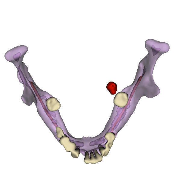
Example 1: Sialolith Mimicking Periapical Radiopacity
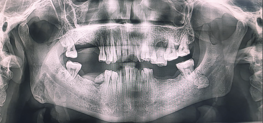
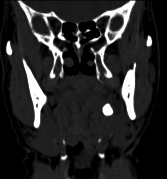
Consider a panoramic radiograph (OPG) displaying a well-defined radiopacity near the periapical region of tooth 38. At first glance, this could be misinterpreted as a dental pathology—perhaps a periapical lesion, cyst, or odontoma. However, when a CT scan is performed, the radiopacity is revealed to be outside the mandibular bone, located within the region of the submandibular gland. The correct diagnosis in this scenario is submandibular gland sialolithiasis, not a dental lesion.
Coronal CT sections may also unveil additional findings, such as calcifications in the neck region. In some cases, these represent Type IV stylohyoid ligament calcifications—a distant calcification type that can be visualized and classified using diagnostic 3D models.
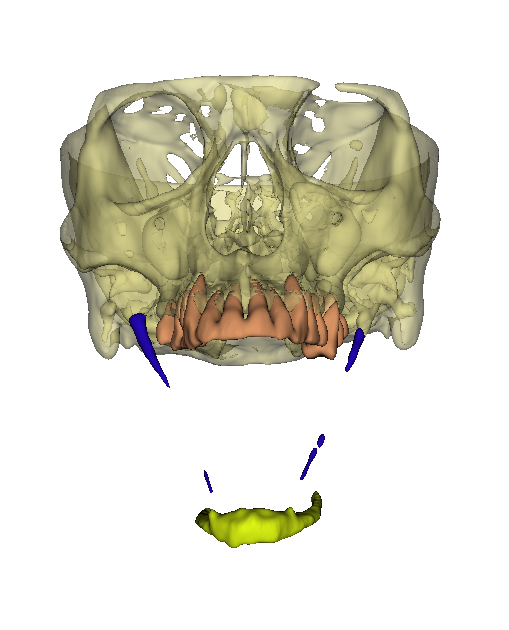
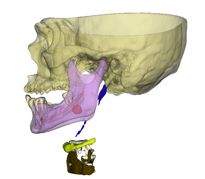
Example 2: Implant Placement and Buccal Cortical Exposure
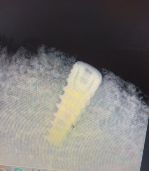
Implantology presents another classic 2D paradox. A post-operative radiograph may show a dental implant that appears perfectly positioned within the bone. The clinician might be satisfied with the placement, especially after achieving the desired torque during insertion. Yet, a CBCT scan may reveal a different story: the implant may penetrate the buccal cortical plate, with partial thread exposure. This exposure makes the implant susceptible to infections and can greatly reduce its longevity—a critical detail often missed in 2D imaging.
More Instances of the 2D Paradox
Dental radiology is rife with cases where 2D images misrepresent the location, extent, or even the nature of pathologies:
- Odontogenic cysts appearing smaller or misplaced.
- Impacted teeth with unexpected angulations.
- Foreign bodies mimicking intra-osseous lesions.
Why 3D Diagnostic Modelling Is Essential?
Diagnostic 3D modelling refers to the process of using advanced imaging modalities—such as Cone Beam Computed Tomography (CBCT) and Computed Tomography (CT)—to create detailed three-dimensional representations of oral and maxillofacial structures. Unlike traditional 2D radiographs, these volumetric images allow clinicians to visualize anatomical relationships, localize pathologies, and assess spatial orientation with exceptional accuracy.
With the evolution of AI in radiology, diagnostic 3D modelling has become even more powerful. Modern AI algorithms are now capable of segmenting oral and craniofacial structures with high precision, automating the identification of teeth, nerves, soft tissues, and bone with minimal manual input. Each anatomical part can be color-coded in the 3D model for clarity, offering both clinicians and patients a more intuitive understanding of complex conditions. This technology not only streamlines diagnosis but also improves treatment planning and enhances patient communication, representing a significant leap in dental imaging capabilities.
Diagnostic 3D modelling bridges the gap in accuracy and visualization, offering significant advantages:
- Precise localization of pathology.
- Accurate assessment of relationships between anatomical structures.
- Clear depiction for clinicians and better communication with patients.
- Improved treatment planning leading to superior outcomes.
Conclusion
The 2D paradox in dental radiology exemplifies the potential pitfalls of relying exclusively on 2D imaging. Embracing 3D diagnostic modalities such as CBCT and CT scans is no longer optional—it’s a necessity. These technologies provide confirmatory diagnoses, empower better treatment planning, and ultimately enhance patient care by revealing the true nature of dental and maxillofacial conditions.

With over a decade of experience in dentistry, I specialize in diagnosing a wide range of oral diseases and have developed a deep passion for dental imaging. My interest in CBCT reporting and digital implantology led me to become a pioneer in the concept of diagnostic 3D modelling. Throughout my career, I have supported and educated numerous dentists in mastering CBCT interpretation, virtual implant planning, and the latest advancements in 3D printing for dental applications. Read more about me on the “about me” page.
- Steiner’s Analysis – Free online Cephalometry - November 23, 2025
- A Complete guide to Grayscale values in CT & CBCT - November 15, 2025
- Struggling to get your old PC to run new Radiology software smoothly? Here’s a trick with SSD that might save you. - November 14, 2025