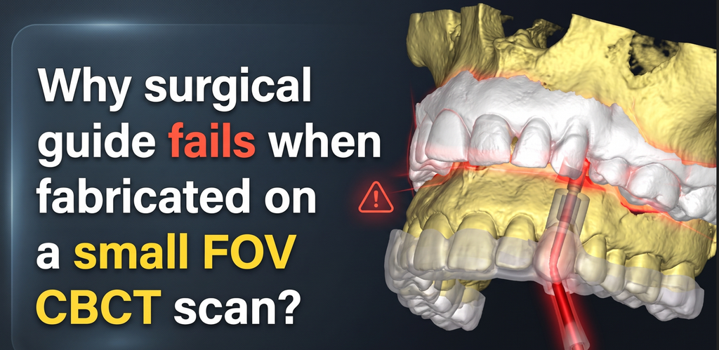
The foundation of any successful guided implant surgery is the precise alignment of the patient’s bone data with their surface anatomy. But a common trap catches many clinicians off guard: combining a limited CBCT scan with a full-arch intraoral scan (IOS). The resulting surgical guide might seat perfectly on the teeth, but the drill trajectory will lead you straight into an error.
Here is exactly why this happens and how you can fix it in your planning software.
What Are Small FOV CBCT Scans?
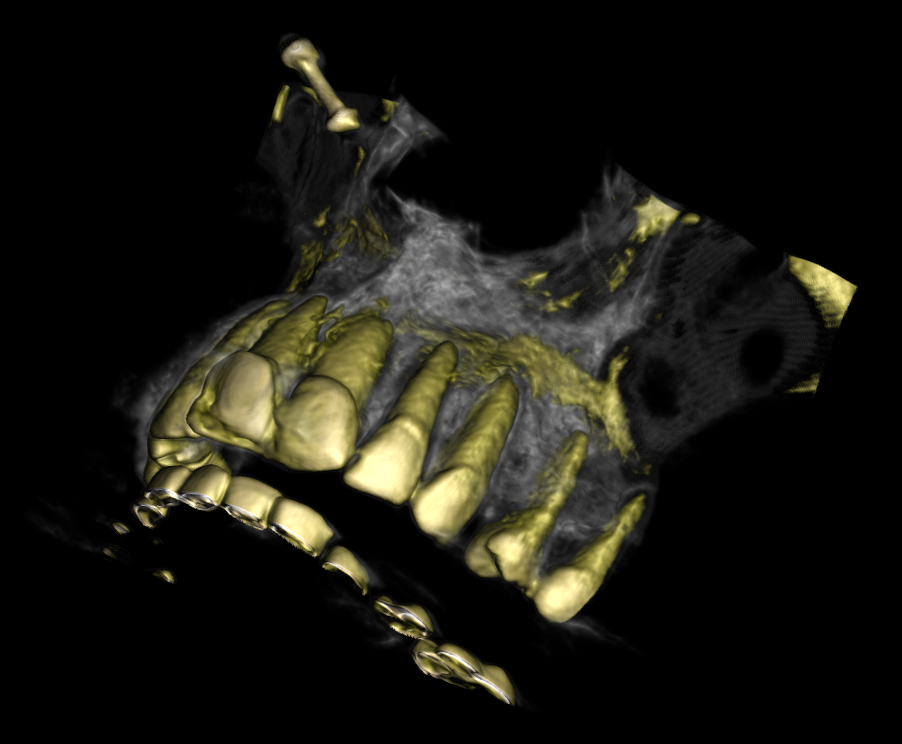
Small Field of View (FOV) CBCT scans typically capture a restricted volume, such as 5×5 cm or 8×5 cm. Clinicians often prefer them for single-implant cases because they offer high spatial resolution with significantly lower radiation exposure to the patient. A small FOV might capture just a single quadrant or a semi-arch (like canine to canine), omitting the posterior regions and the opposing arch entirely. While excellent for diagnostics, this limited data presents a unique mechanical challenge when it is time to design a surgical guide.
How CBCT and IOS Superimpose Using ICP
To design a guide, the software must stitch the DICOM data (CBCT) with the STL/PLY data (IOS). This is done using the Iterative Closest Point (ICP) algorithm. ICP works by treating both scans as a cloud of 3D points. The algorithm calculates the distances between the points on the IOS and the corresponding points on the CBCT tooth surfaces. It then iteratively shifts and rotates the IOS until the overall mathematical distance between the two surfaces is as close to zero as possible, locking them into a “best-fit” alignment.
The Role of Modern AI in Superimposition
Modern implant planning software uses AI to speed up this process. Instead of manually picking three or four corresponding points on both scans, AI automatically segments the teeth from the CBCT bone data, converting them into a high-contrast 3D mesh. The AI then recognizes the shape of the clinical crowns on both datasets and performs an automatic initial alignment. Once the AI brings the scans close together, the ICP algorithm takes over to refine the micro-alignment.
The Core Problem: Why Full-Arch IOS Fails on Small FOV Scans
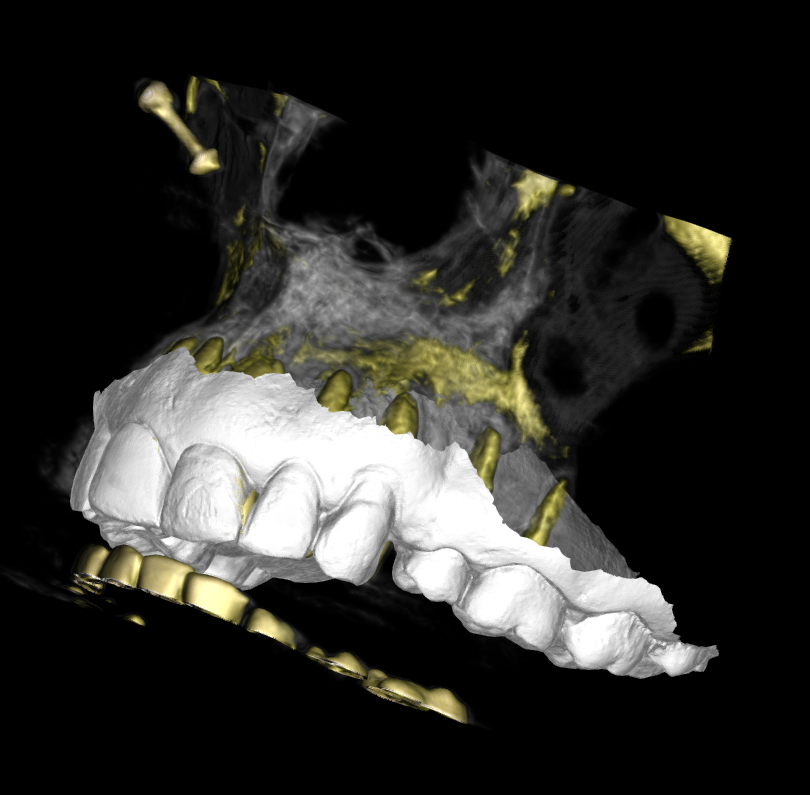
The superimposition breaks down when you feed the algorithm mismatched datasets. If your CBCT only shows teeth from the right first premolar to the left first premolar, but your IOS contains the entire maxillary arch including the molars, the software gets confused.
The ICP algorithm aggressively tries to find a match for those posterior molars present in the IOS. Since those molars do not exist in the small FOV CBCT, the software forces a compromised mathematical average. It tilts, shifts, or slightly rotates the IOS to account for the missing data.
To the naked eye, the front teeth might look reasonably aligned, but the overall mesh is deviated. Because the guide is fabricated based on the IOS, it will fit snugly in the patient’s mouth. However, the implant planning was done on the underlying shifted CBCT bone. When you drill, your osteotomy will follow the software’s invisible error, placing the implant in a deviated, potentially dangerous position.
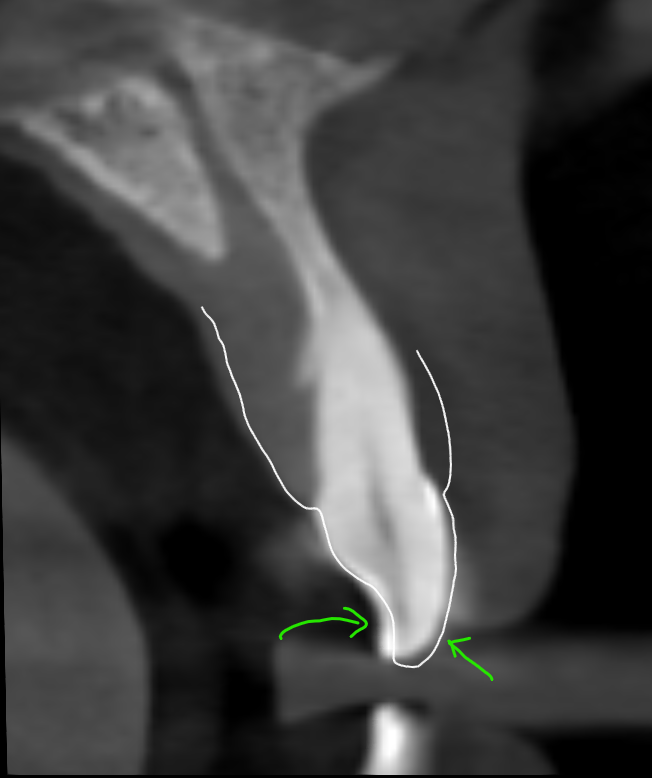
How to Rectify the Alignment Error
The solution is dataset equalization. You must give the ICP algorithm a 1:1 match.
Before you run the superimposition tool in your software, take a moment to digitally crop your intraoral scan. Evaluate your small FOV CBCT and count the exact number of teeth visible. Identify the terminal teeth on both sides of the scan (for example, tooth #14 to #24).
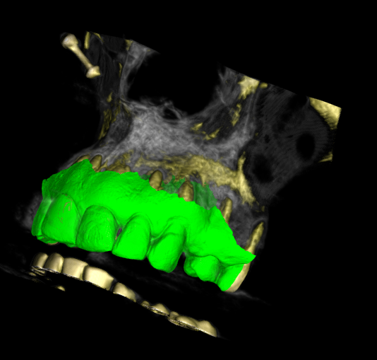
Once you have identified the boundaries, open your IOS editing tool and cut away everything distal to #14 and #24. By digitally amputating the unrepresented posterior teeth from the intraoral scan, you remove the excess data that derails the ICP algorithm.
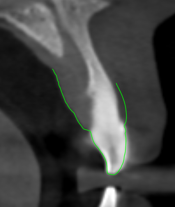
When you superimpose this accurately cropped IOS onto the small FOV CBCT, the AI and ICP algorithms have exactly the same anatomical landmarks to work with. The software no longer struggles to calculate missing data, resulting in a flawless, error-free superimposition. Taking two minutes to trim your scan ensures your surgical guide actually guides your drill exactly where you planned it.
Conclusion
Digital implantology is only as precise as your initial data alignment. While small FOV CBCT scans are excellent for minimizing patient radiation and capturing high-resolution localized details, they require a strict workflow adjustment during software planning. Attempting to stitch a full-arch intraoral scan over a limited CBCT volume is a guaranteed way to introduce invisible, algorithm-driven deviations into your surgical guide.
By taking just a few extra moments to crop your IOS to the exact dental boundaries of your CBCT, you give the ICP algorithm the 1:1 anatomical match it needs to function perfectly. This simple digital adjustment is a non-negotiable rule for small FOV cases. Master this step, and you ensure that the exact trajectory you plan on your screen translates flawlessly to the surgical site.

A passionate dental educator with more than 13 years of academic experience, I am dedicated to making complex imaging feel simple by always starting from the basics. Technology is my playground—specializing in CBCT reporting, digital implantology, 3D modelling, and dental 3D printing innovations. Through my writing and teaching, I love empowering clinicians to bridge the gap between advanced digital workflows and everyday clinical practice.
Read more about me on the “about me” page.
- Why surgical guide fails when fabricated on a small FOV CBCT scan? Read this before you fabricate one. - November 12, 2026
- Are Intraoral Scans Actually Distortion-Free? Know this before you buy one - November 1, 2026
- Decoding TMJ Pain: Why Open-Mouth CBCT Views Are a Game-Changer in TMJ Diagnosis - October 30, 2026