Recently i have come across an interesting article on Dedicated Dental MRI by Jennifer Christensen and Rubens Spin-Neto. The much needed advancement in Dental Imaging.
Dentistry has long relied on X-rays, panoramic radiographs, and Cone Beam Computed Tomography (CBCT) to visualize teeth and jaw structures. While these tools are effective, they come with a notable trade-off: ionizing radiation.
Enter Dental MRI (specifically, Dental-Dedicated MRI or ddMRI)—a revolutionary shift in oral and maxillofacial diagnostics that offers unprecedented soft-tissue visualization completely free of radiation.
In this post, we will break down what dental MRI is, who created it, how it works, and how it is poised to transform the future of dental care.
What is Dental MRI?
Dental MRI is a specialized magnetic resonance imaging application optimized for visualizing the teeth, jawbones, gums, nerves, and surrounding soft tissues. Unlike conventional dental X-rays or CT scans that excel primarily at capturing hard bone structures, dental MRI shines by providing crystal-clear images of soft tissues, nerves, blood vessels, and early inflammatory changes—all without exposing the patient to potentially harmful ionizing radiation.
2. Who Made It and Why?
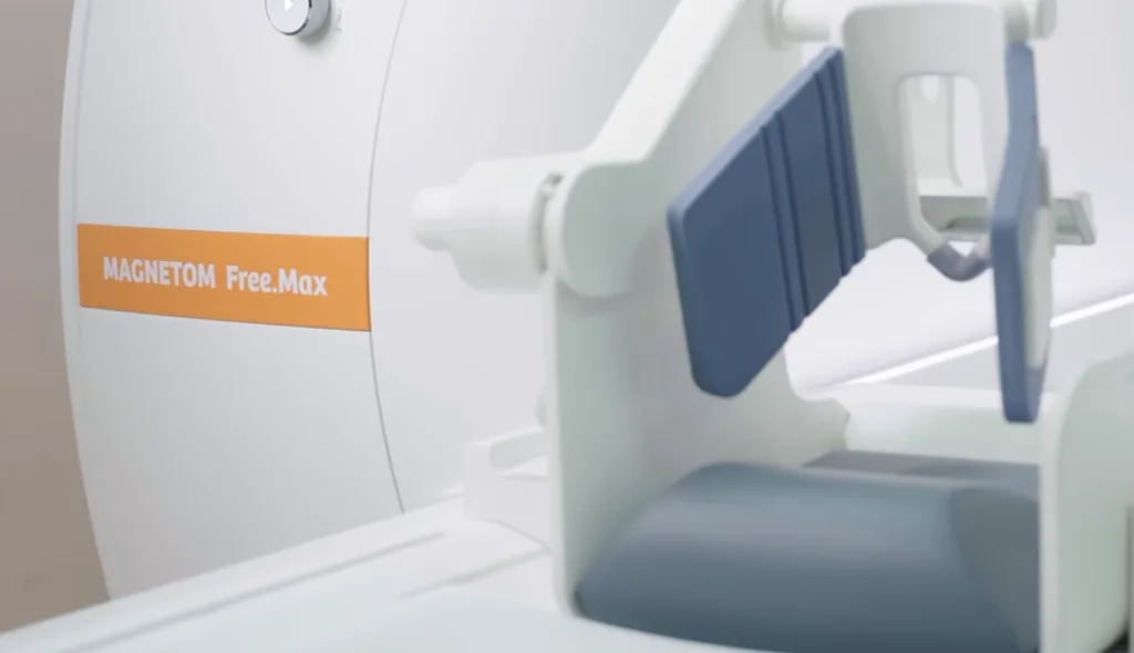
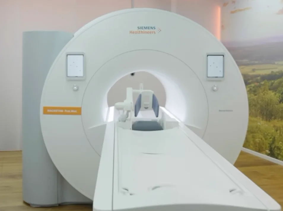
The breakthrough commercialization of dental-dedicated MRI is spearheaded by medical technology leaders like Siemens Healthineers (such as the MAGNETOM Free.Max Dental Edition), often in close collaboration with academic dental research institutions and clinical partners.
Why was it created?
- The Radiation Safety Imperative: Conventional imaging methods like CBCT use X-rays, which carry cumulative DNA-damaging risks. Dental MRI was developed to make the “As Low As Reasonably Achievable” (ALARA) principle obsolete by eliminating radiation entirely.
- The Soft-Tissue Blind Spot: Traditional dental tools are poor at imaging fine soft-tissue structures like the interior alveolar nerve or early-stage soft-tissue lesions. Clinicians needed a way to view nerves, pulp, and periodontal ligaments with high fidelity.
How is Dental MRI Different from Conventional MRI?
While a standard whole-body medical MRI can theoretically scan the head, they are rarely used for routine dental checks due to severe technical limitations. Dental MRI differs in several key ways:
- Field Strength Optimization: Standard clinical MRIs operate at high field strengths (1.5 Tesla or 3.0 Tesla). Dental-dedicated MRI systems often utilize an optimized 0.55 Tesla “High-V” field strength, which significantly reduces metal artifacts from dental fillings and crowns.
- Dedicated Coils and Sequences: Dental MRIs use specialized radiofrequency coils designed specifically to conform tightly to the oral cavity, delivering high signal-to-noise ratios (SNR) specifically for the jaw region.
Software Update vs. Capturing Mechanism: What Changed?
Is dental MRI just a software patch, or is the hardware different? The capturing mechanism (hardware) has fundamentally changed.
- The Hardware Revolution: You cannot simply run a standard whole-body MRI scanner with new software to perform effective dental imaging. Dental MRI requires specialized, high-density radiofrequency receiver coils designed to fit closely around the mouth and jaw, alongside customized gradient systems to handle tiny, high-resolution voxels in a region prone to magnetic susceptibility artifacts.
- The Software Evolution: Alongside the hardware, cutting-edge software—such as AI-powered reconstruction algorithms, specialized ultra-short echo time (UTE) sequences, and deep learning image acceleration—plays a vital role in cleaning up images and cutting down scan times.
Key Clinical Applications
Dental MRI unlocks diagnostic capabilities that were previously difficult or impossible to achieve with standard X-rays:
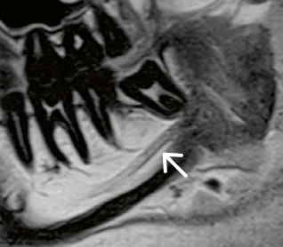
Third Molar (Wisdom Tooth) Surgery: Precise mapping of the inferior alveolar nerve inside the mandibular canal, drastically reducing the risk of permanent nerve damage and numbness.
Dental Implant Planning: Accurate visualization of the gingiva, bone architecture, and nerve pathways to ensure flawless implant positioning.
Temporomandibular Joint (TMJ) Disorders: Unrivaled capability to diagnose articular disc displacements, micro-tears, and joint inflammation in the TMJ.
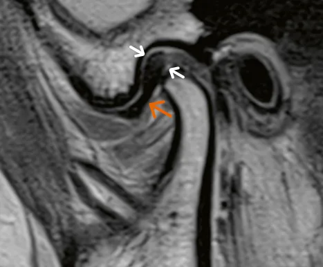
Endodontics and Periodontics: Early detection of root canal inflammation, periapical lesions, and periodontal soft-tissue pockets without radiation.
How Dental MRI Will Change the Future of Dental Imaging
The integration of dental MRI into mainstream dentistry represents a paradigm shift from hard-tissue-only diagnostics to comprehensive holistic diagnostics.
As technology advances—and as manufacturers refine hybrid workflows that merge the bone-imaging precision of CT with the soft-tissue mastery of MRI—dentists will be able to perform fully integrated, zero-radiation treatment planning. For patients, this means safer check-ups, highly personalized oral surgeries, and earlier detection of pathologies long before they manifest structurally on conventional X-rays.
Dental MRI is no longer just a futuristic concept; it is actively setting a new gold standard for patient safety and diagnostic accuracy in modern dentistry.

With over a decade of experience in dentistry, I specialize in diagnosing a wide range of oral diseases and have developed a deep passion for dental imaging. My interest in CBCT reporting and digital implantology led me to become a pioneer in the concept of diagnostic 3D modelling. Throughout my career, I have supported and educated numerous dentists in mastering CBCT interpretation, virtual implant planning, and the latest advancements in 3D printing for dental applications. Read more about me on the “about me” page.
- What is Dental MRI? The Future of Radiation-Free Oral Imaging - October 25, 2026
- The Dual-Kit Dilemma: Why Fragmented Guided Implant Systems Are Losing Ground to Single-Kit Solutions - October 24, 2026
- A Complete guide to Grayscale values in CT & CBCT - November 15, 2025