What Are Dentigerous Cysts in Maxillary Teeth?
A dentigerous cyst (also called a follicular cyst) is a type of developmental odontogenic cyst that forms around the crown of an unerupted or impacted tooth. It develops when fluid accumulates between the reduced enamel epithelium and the crown of the tooth, creating a cystic cavity.
In the maxillary (upper jaw) region, dentigerous cysts are most commonly associated with impacted canines and third molars (wisdom teeth). Because of the limited bone space and proximity to the maxillary sinus, these cysts can sometimes expand and cause displacement of adjacent structures.
Key Features
- Location: Commonly around impacted maxillary canines or third molars.
- Radiographic Appearance: A well-defined, unilocular radiolucency attached at the cementoenamel junction (CEJ) of an unerupted tooth.
- Symptoms: Usually asymptomatic initially, but may cause painless swelling, tooth displacement, or sinus expansion as it grows.
- Complications: Large cysts can cause root resorption of adjacent teeth or displacement of the maxillary sinus.
- Diagnosis: Typically confirmed with CBCT imaging for 3D visualization of the cyst, the associated tooth, and nearby structures.
- Treatment: Surgical enucleation of the cyst and removal of the associated tooth, followed by histopathological confirmation.
Case Overview
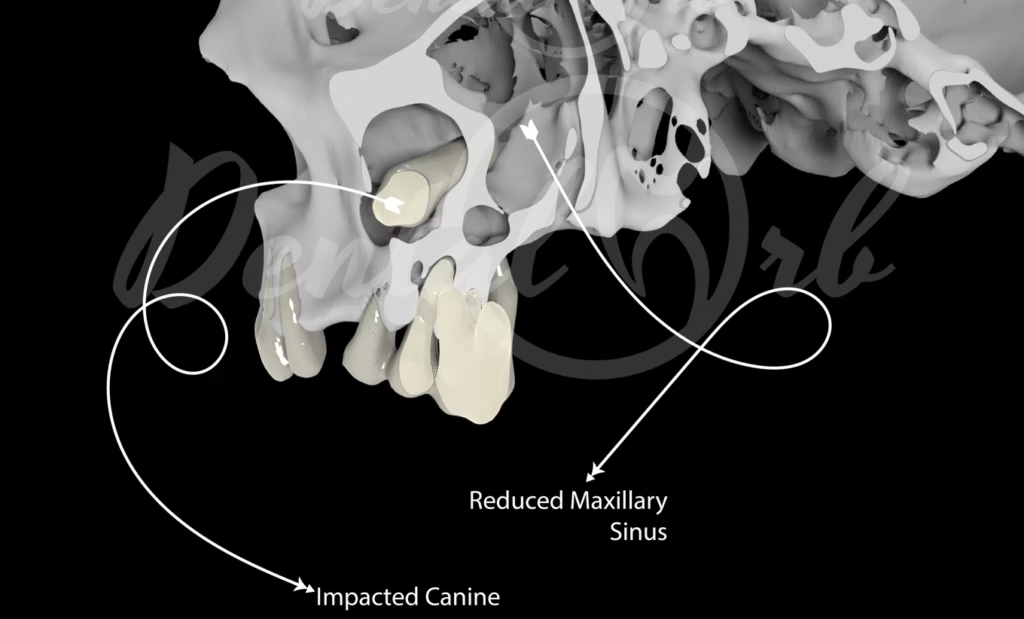
This is an interesting case of a dentigerous cyst developing around an impacted maxillary canine, where the lesion gradually expanded into the surrounding spaces. Over time, the cyst caused a significant displacement of the maxillary sinus, providing a fascinating insight into the anatomical relationships of the region.
Clinical Findings
The impacted canine was completely embedded in bone, with its crown portion facing anteriorly and lying flat above the existing dentition. Unfortunately, a dentigerous cyst formed around the crown portion of this impacted tooth.
What makes this case unique is the direction of cyst expansion — it displaced the maxillary sinus posteriorly and superiorly, yet there were no clinical signs of sinusitis. This unusual presentation highlights how expansile cystic lesions can alter adjacent structures without necessarily causing inflammatory symptoms.
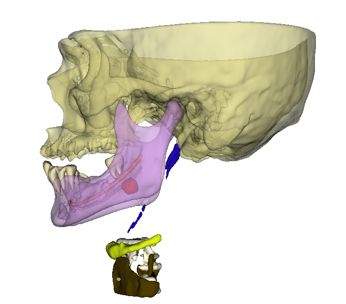
3D Diagnostic Modeling
To better understand the complex spatial relationships, a diagnostic 3D model was created from the CBCT data. This model allowed for precise visualization of the impacted tooth, the cyst surrounding it, and the adjacent sinus anatomy.
By sectioning the skull in the virtual model, we were able to appreciate the internal structures and the extent of displacement in remarkable detail. Such 3D visualization not only enhances diagnostic accuracy but also serves as an excellent educational and surgical planning tool.
Conclusion
This case beautifully demonstrates how diagnostic 3D modeling can transform our understanding of pathologies like dentigerous cysts. The ability to visualize internal anatomy in three dimensions helps clinicians appreciate the true extent and impact of lesions, leading to more informed treatment planning.
Below is a rendered video from the 3D model that capture the condition in stunning detail — we hope you enjoy exploring them as much as we did creating them.

With over a decade of experience in dentistry, I specialize in diagnosing a wide range of oral diseases and have developed a deep passion for dental imaging. My interest in CBCT reporting and digital implantology led me to become a pioneer in the concept of diagnostic 3D modelling. Throughout my career, I have supported and educated numerous dentists in mastering CBCT interpretation, virtual implant planning, and the latest advancements in 3D printing for dental applications. Read more about me on the “about me” page.
- Steiner’s Analysis – Free online Cephalometry - November 23, 2025
- A Complete guide to Grayscale values in CT & CBCT - November 15, 2025
- Struggling to get your old PC to run new Radiology software smoothly? Here’s a trick with SSD that might save you. - November 14, 2025